"Life-Changing… Nothing Short of Miraculous" — Google Reviews
The Why Behind It All
Science that explains what you feel and how to get better!
- ADHD
- PTSD
- Traumatic Brain Injury
- Anxiety & Depression
- Cognitive Decline
- Suicidal Thoughts
- Performance Optimization
- Autism
- Postpartum
- Concussions
- Parkinson’s
- Multiple Sclerosis
- Brain Fog
Proven across 20,000+ brains, the GBI Quant360 Functional Analysis goes beyond traditional testing to show why you feel the way you do and what to do about it. See the real data behind your brain and nervous system, and get a personalized plan to restore clarity, regain control, and finally feel like yourself again.
You’ve done the tests. You’ve tried the pills. You’ve waited for answers that never came. It’s time for something different.
People from age 5 to 100 come to Genesis Brain Institute because they’ve followed the rules. They’ve taken the pills, done the checkups, and waited for results, but still don’t feel like themselves. They’re done with surface-level testing and temporary fixes. They want real answers that explain what’s actually happening and measurable steps to improve it.
The Quant360 Functional Analysis is the first step toward:
Improved focus & attention
Reduced stress and anxiety
Sharper thinking and memory
A brighter, more joyful outlook
Greater mental clarity
Better brain-body coordination
This is Healing

This is ADHD.

This is Cognitive Decline.
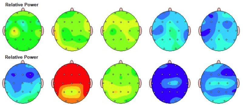
This is Anxiety.
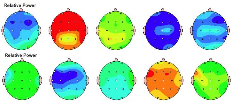
This is Trauma / PTSD.

This is Depression.

This is OCD.

This is a TBI.

Answers. Recovery. Potential.
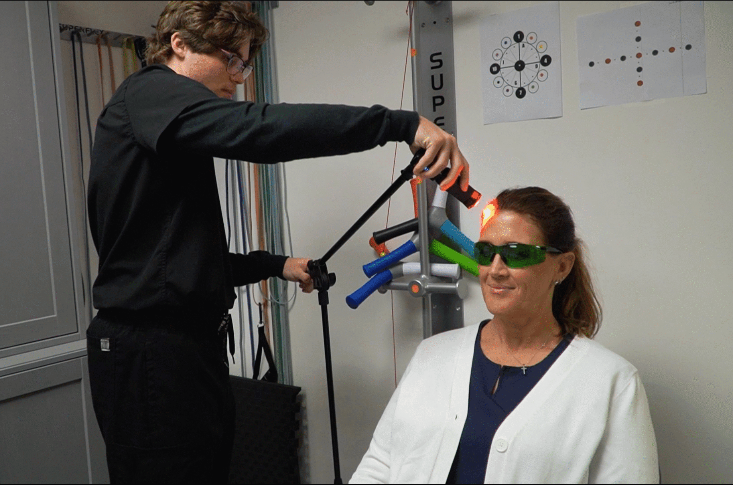
Looking for real answers about your brain health?
People ages 5 to 100 come to Genesis Brain Institute because they want more than a label — they want solutions. The GBI Quant360 Functional Analysis is the first step toward understanding what’s really happening in your brain and body, and how to improve it.
This is the first step towards:
- Improved focus & attention
- Reduced stress and anxiety
- Sharper thinking and memory
- A brighter, more joyful outlook
- Greater mental clarity
- Better brain-body coordination
The GBI Quant360 Functional Analysis
Our elite team of brain and nervous system specialists — led by licensed doctors and clinical experts — take you through a 2–3 hour, multi-step process to uncover what’s really happening inside your brain, and why past treatments may have missed it. From qEEG Brain Mapping to balance, eye movement, cognitive testing, and more, every step gives us hard data to create a plan that’s tailored to you — not your symptoms.
Finally Answers and a Plan for challenges like:
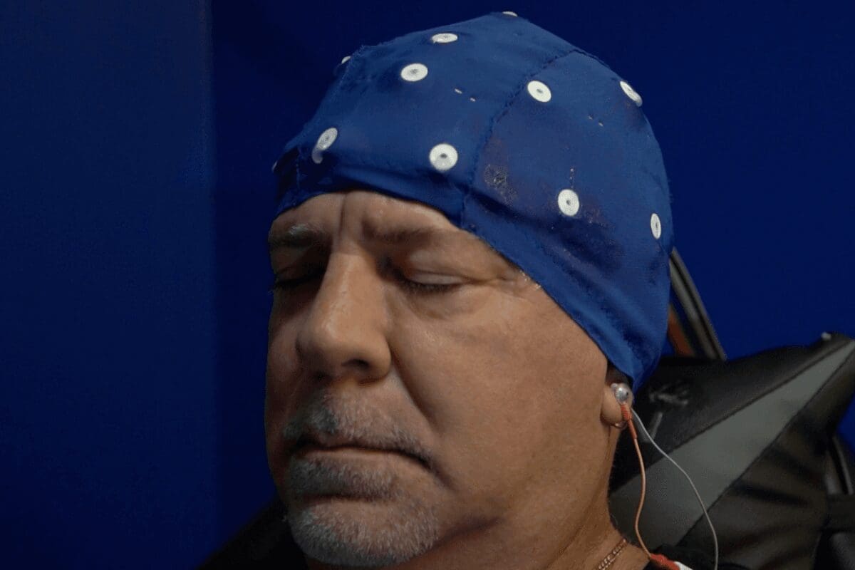
When You Measure Against 20,000 Brains, Patterns Emerge. Answers Appear.
Our technology is built on one of the largest brain databases in the world, backed by 40+ years of research and over 200 published studies — combined with our full suite of science-backed analyses and tests to detect patterns and problems other approaches miss.
Posted onTrustindex verifies that the original source of the review is Google. If the initial consult is any indication of how treatment is going to go for my son struggling with some mental health issues, I am VERY encouraged. Dr K seems very competent, credentialed, knowledgeable, and her bedside manner is awesome!Posted onTrustindex verifies that the original source of the review is Google. I'm 67 and I have never been to a medical institution with more professionalism than Genesis Brain Institute. Everyone there makes you feel so cared about. Thank you Everyone.Posted onTrustindex verifies that the original source of the review is Google. fantastic staff and talented drs for treatmentPosted onTrustindex verifies that the original source of the review is Google. It was nice experience because they care about you and I needed that and I would recommend anyone tho Genesis Brain institute they really helped me to be honest with you I didn't think that I was going to be able to do it and I'm glad I did like I said it was a very nice experiencePosted onTrustindex verifies that the original source of the review is Google. After having a QEEG and receiving treatment I notice a night and day difference in my energy levels, quality of sleep and have an overall reduction in anxiety. Infected with Lyme disease and living with chronic stress for extended periods of time I knew I needed to assess and improve my cognitive functioning. I couldn’t be more pleased with my results and I am confident that I will continue to improve with the guidance of GBI. Thank you to Dr. K and the team 🙂Posted onTrustindex verifies that the original source of the review is Google. I had an amazing experience at the Genesis Brain Institute! Their personalized treatments truly made a difference in how I feel. After undergoing TMS therapy, I felt lighter, more hopeful, and genuinely more like myself. The staff is incredibly supportive and knowledgeable. I highly recommend them!Posted onTrustindex verifies that the original source of the review is Google. I am currently in treatment with the GBI due to a head injury from a car accident. The team has been very helpful from the beginning from scheduling to treatment. I am already seeing progress. Conner and crew, who do my weekly therapy, provide a comfortable environment with lots of encouragement. Dr. K is wonderful to work with as well.Posted onTrustindex verifies that the original source of the review is Google. I have experienced nothing short of the friendliness, feeling comfortable , caring and support that has been shown to me throughout my process thus far..the staff is great!
The GBI Quant360 Functional Analysis is the First Choice for People Who Want Answers
Who We’re Trusted By:
The Proven Path to Real Answers, Real Healing, Hope and a Better Brain—Without Guesswork or Meds
- Why your child still struggles to focus or manage emotions
- Why you can’t remember names, tasks, or where your keys went
- Why anxiety, brain fog, or PTSD symptoms won’t let up
- Why you’re not performing at the level you know you could be
And most importantly, what you can do about it—based on real data from your brain.
At GBI, our premier diagnostics show what parts of your brain are running too fast, too slow and just right—so we can treat the condition without meds… or help you unlock peak performance and gain an edge at school, work, home and life!

Discover What’s Really Going On In Your Brain
This Isn’t Just for One Type of Person. It’s for You.
Whether you’re a mom desperate to help your child focus or regulate emotions, an executive chasing clarity and peak energy, a veteran or first responder battling anxiety or PTSD, or a senior noticing your memory isn’t what it used to be—this is your next step.
You don’t have to keep guessing. You don’t have to accept “this is just the way it is.” You don’t have to try one more thing that doesn’t work.
At Genesis Brain Institute, our diagnostics uncover what others miss. If something feels off—we find out why. If you’re doing “fine”—we show you how to do even better. If you’ve tried everything and nothing has worked—this is where your breakthrough begins.
Why This Brain Scanning Is Different
This isn’t a quick quiz or questionnaire. This is a 3-hour, physician-led series of advanced brain tests looking at your Brain Condition—supported by a powerful in-house team that includes:
- A Board-Certified Neurosurgeon
- A Clinical Neuroscientist & Functional Neurologist
- A Board-Certified Pediatric Physician
- A Board-Certified Mental Health Nurse Practitioner
- A Licensed Mental Health Counselor
It’s a full-spectrum brain and body assessment that helps you:
- Get to the root of anxiety, depression, ADHD, TBI symptoms, cognitive decline, fatigue, and more
- Uncover how your brain is functioning, not just how you feel
- See the exact areas where your brain is underperforming (or overworking)
- Know where to treat—and how to track progress
- Identify how to reach your next level if you’re already doing well

Learn More About Each Diagnostic Test
Want to understand the science of why our diagnostics catch what others miss? Here’s how we uncover what’s really going on in your brain:
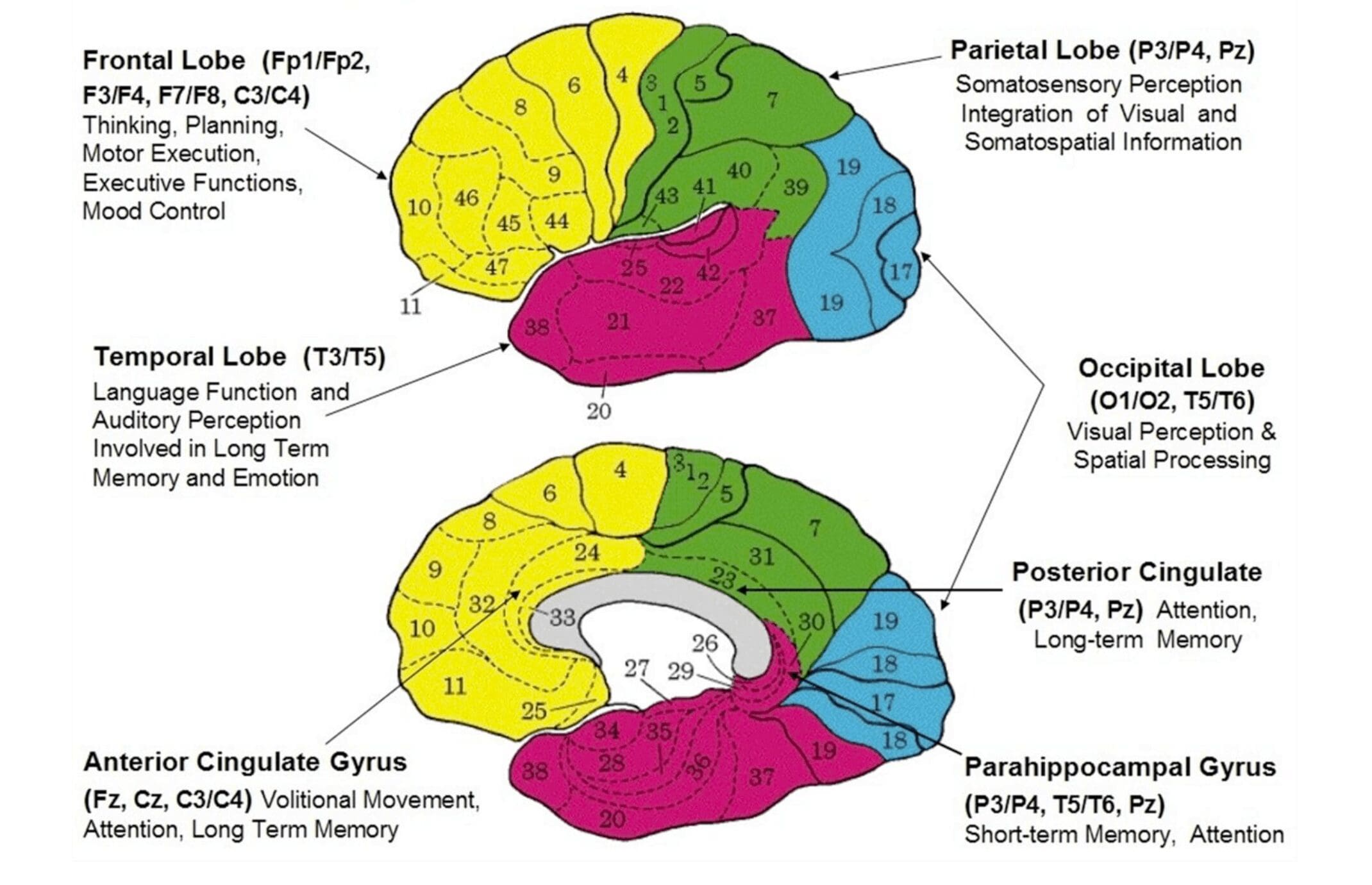
qEEG Brain Mapping
This is our starting point. It shows us what areas of your brain are overactive, underactive, or working just right—providing the GPS to design your treatment path.
Videonystagmography (VNG) & Balance Testing
Helps detect issues in your vestibular system (inner ear and brain balance centers) that may be contributing to dizziness, instability, or coordination issues.
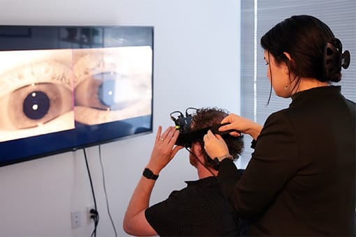
Oculomotor Assessments
Tracks how your eyes move and process information—key for anyone dealing with brain fog, TBI, or performance issues.
Cognitive Performance Testing
Measures speed, memory, focus, and how efficiently your brain makes decisions.
MRI Review (if needed)
While our focus is on function, we also review structural imaging like MRIs if our Neurosurgeon requests this, giving our team another layer of insight into what may be affecting your brain health.
Consultation with Functional Neurologist
We take everything we've learned from your diagnostics and sit down one-on-one to explain your results, answer your questions, and design a fully customized treatment protocol for your brain.
Each diagnostic is non-invasive, deeply informative, and reviewed by our full interdisciplinary team. It’s not just about what’s happening—but why it’s happening and what to do about it.. Each result is reviewed by multiple members of our expert team. Each one contributes to a personalized roadmap to healing and performance.
Which One Are You?
The Overwhelmed Parent
"I just want to help my child thrive without more meds." → We identify what your child’s brain actually needs—and create a plan.
The Burnt-Out Professional
"My mind is always running, but my energy is fading." → We show you where your cognitive systems are under stress and how to restore clarity, memory, and drive.
The Stuck but Strong Adult
"I’ve been to therapy. I’ve taken the meds. I still feel off." → We uncover root-level causes of mental health struggles and build a non-medication path forward.
The Aging Achiever
"I’m slipping. I want to stay sharp for my family and future." → We detect early signs of decline and create a brain health plan that protects your tomorrow.
The High Performer
"I’m doing well—but I know I could do more." → We find the micro-imbalances that may be blocking your next-level speed, energy, and resilience.
The Misunderstood Survivor
"It was ‘just a concussion’—but I haven’t felt the same since." → Whether it’s from a concussion or TBI, we measure the unseen damage and build a personalized recovery plan to restore clarity, stability, and your sense of self.
Your Story Matters. Your Brain Deserves This.
When was the last time someone looked at your full story and your full brain function? Looking at each specific area of your brain?
Most of our clients say, “This is the first time I feel seen, heard, and finally understood.”
Because our diagnostics don’t just show us what’s wrong—they help us plan what to do right. This is the beginning of hope.
Whether you’re hurting or just hungry to reach the next level—it all starts here.
Genesis Brain Institute: The Team Behind the Science
What sets us apart isn’t just the equipment—it’s the people behind it.
Every diagnostic is reviewed by a cross-disciplinary team with decades of combined experience in:
- Neurosurgery
- Functional Neurology
- Pediatrics
- Cognitive and Emotional Disorders
- Elite Athlete Optimization
- TBI and Concussion Recovery
This is not a franchise. This is not cookie-cutter. This is everything under one roof. No more running from clinic to clinic, collecting opinions from doctors who don’t talk to each other or rushing into prescriptions that mask symptoms instead of fixing the problem. This is personalized, physician-directed care that targets the root cause—so you can heal, improve, and perform at your best without relying on medication.
If you’re in Tampa and looking for real answers for your mind, your child, or your performance—this is where the journey begins.
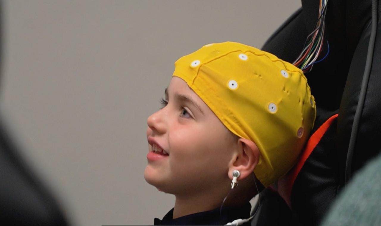
Quantitative Electroencephalogram (qEEG)
Electroencephalograms (EEGs) have been used for decades to help neurologists diagnose epilepsy,

Video Oculography / Nystagmography
Eye movements have been keenly reviewed by medical scientists to relate different brain networks to specific eye movements.
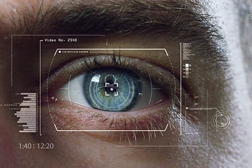
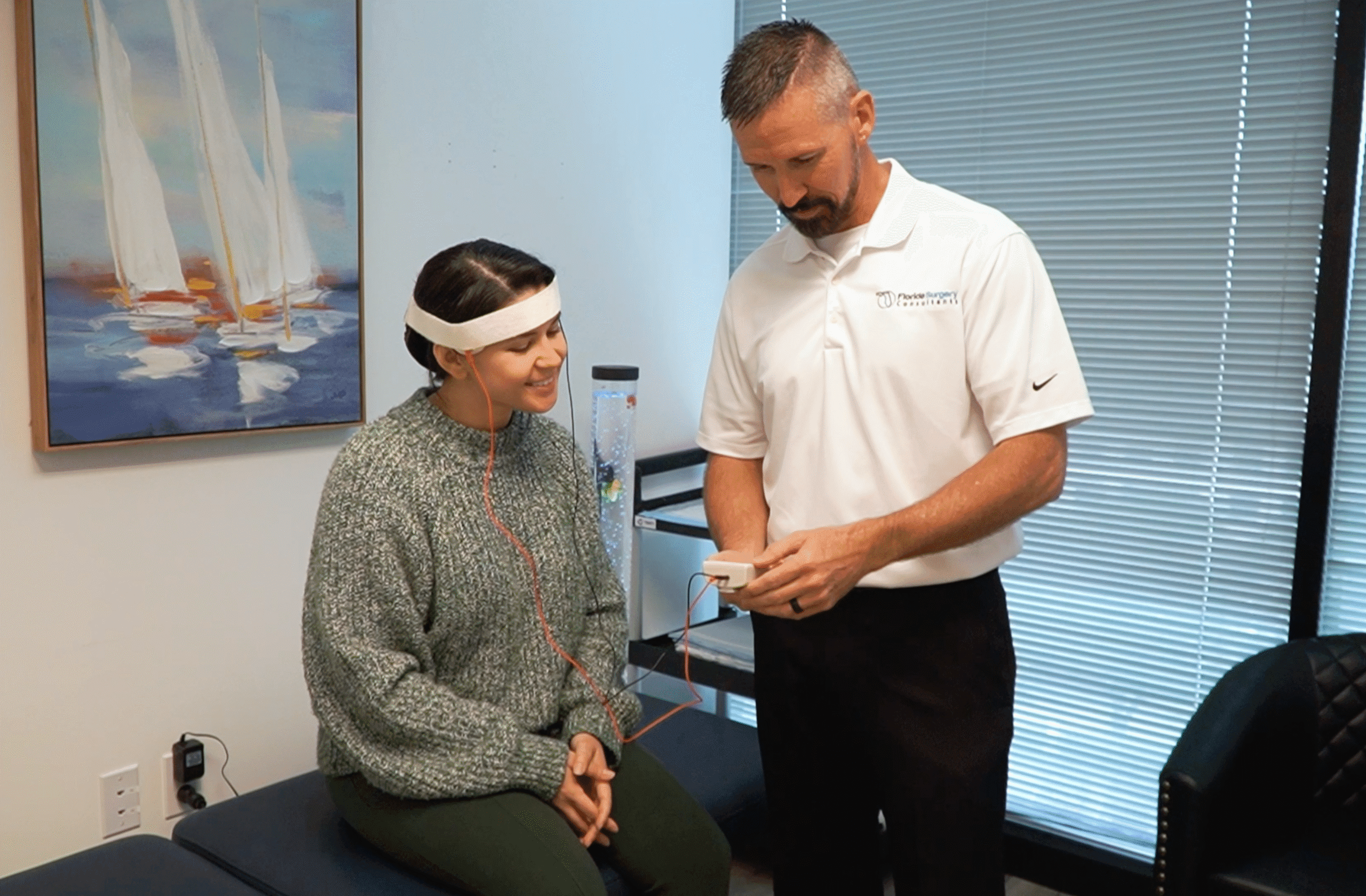
Pupillometry via Reflex Pro
Pupillometry stands as a validated clinical instrument, offering a swift and objective biomarker for autonomic responses.
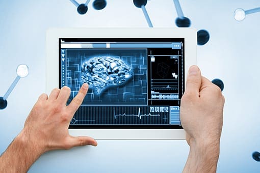
Cognitive Performance Testing
Our clinical team uses objective cognitive testing to determine which cognitive functions may be affected and to monitor improvements
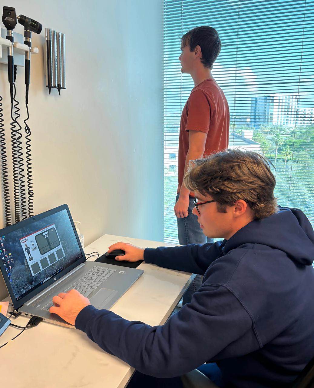
Balance Assessment
Balance is more than just standing upright. At GBI, we utilize computerized posturography with a modified clinical test of sensory integration

Brain MRI Review
Magnetic resonance imaging is a medical imaging technique used to create pictures of the brain using strong magnetic fields


